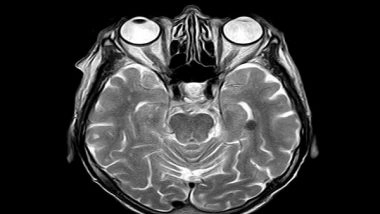
Stockholm [Sweden], December 23 (ANI): Brain age is an MRI-derived estimate of brain tissue loss that has a similar pattern to aging-related atrophy. White matter hyperintensities (WMHs) are neuroimaging markers of small vessel disease and may represent subtle signs of brain compromise.
A new research paper has been published in the journal Aging, titled 'White matter hyperintensity load is associated with premature brain aging'.
In this new study, researchers Natalie Busby, Sarah Newman-Norlund, Sara Sayers, Roger Newman-Norlund, Sarah Wilson, Samaneh Nemati, Chris Rorden, Janina Wilmskoetter, Nicholas Riccardi, Rebecca Roth, Julius Fridriksson, and Leonardo Bonilha from the University of South Carolina, Medical University of South Carolina and Emory University tested the hypothesis that WMHs are independently associated with premature brain age in an original aging cohort.
"We hypothesized that a higher WMH load is linearly associated with premature brain aging controlling for chronological age."
Brain age was calculated using machine learning on whole-brain tissue estimates from T1-weighted images using the BrainAgeR analysis pipeline in 166 healthy adult participants. WMHs were manually delineated on FLAIR images. WMH load was defined as the cumulative volume of WMHs.
A positive difference between estimated brain age and chronological age (BrainGAP) was used as a measure of premature brain aging. Then, partial Pearson correlations between BrainGAP and the volume of WMHs were calculated (accounting for chronological age).
Brain and chronological age were strongly correlated (r(163)=0.932, p
(The above story is verified and authored by ANI staff, ANI is South Asia's leading multimedia news agency with over 100 bureaus in India, South Asia and across the globe. ANI brings the latest news on Politics and Current Affairs in India & around the World, Sports, Health, Fitness, Entertainment, & News. The views appearing in the above post do not reflect the opinions of LatestLY)


 Quickly


